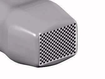
Figure 17: Schematic image of an ultrasound probe for 3D imaging with a matrix array.
The first functional probes based on matrix arrays (fig 17) appeared in 2004. This probe can be controlled in two planes and does not need any mechanical movement. The first systems can record a volume image of the heart in a few beats, but the development is moving toward recording the entire heart in just a single beat. A matrix probe for the heart typically has 2-4000 elements. Both the large number and their high impedance make it impossible to connect them directly to the scanner through a cable. Consequently, the beam formation is split into two stages, an integrated micro beamformer in the probe and a conventional beam former of typically 128 channels in the scanner. The first systems used an analog micro beamformer which reducesdthe number of elements to 128. In addition, the scanner must have parallel beams in order to increase the frame rate (Multi Line Acquisition).
The transmitters in the system must be able to excite the entire probe. The best is to have a configurable switch in the probe. But the alternative is to have a fast connection, for example an array of diodes, so that the elements are connected together for high-level transmitting signals, but not for low-level receiving signals. As the transmitter often operates on a relatively low frequency in harmonic imaging and in Doppler modes, one can connect together 2x2 elements for example. 256 transmitters are then distributed over 1024 elements which will cover 25-50% of the transducer surface. This gives a wider transmitting beam than the receiving beam, which is desirable because it should cover multiple parallel receiver beams.
Figure 17: Schematic image of an ultrasound probe for 3D imaging with a matrix array.