Mechanical waves with oscillations (frequency) above the audible range is called ultrasound. Ultrasound is characterized by:
- Intensity, given by the pressure amplitude
- Frequency, measured in Hertz (Hz). Audible sound is between 20 and 20,000 Hz. In typical medical ultrasound, frequencies between 2 and 10 MHz are used. In intravascular imaging (imaging from the inside of blood vessels), frequencies of up to 30 MHz are used.
- Wavelength, hereafter represented with λ.
The speed of sound (c) is an important property of the medium in which sound propagates. It is 340 m/s in air and about 1500 m/s in water. Tissue is similar to water, and has a speed of sound of around c = 1540 m/s. Since wavelength λ = c/f, the wavelength for medical ultrasound is between 0.15 mm and 0.75 mm. In comparison, the wavelength for audible sound lies in the range 2 cm to 20 m. Ultrasound of the body can be used for imaging up to a depth of about 20 cm. Within this scale, ultrasound has properties which more resemble the properties of visible light than those typically associated with audible sound:
- The sound waves travel in a straight line which can be focused and deflected.
- A sound wave that encounters a medium which has significantly different properties from soft tissue will be nearly completely reflected. Differences in the product of density and sound are the determining factors.
The largest reflection occurs when meeting bone or air. This means that one cannot obtain an image if there is a part of a lung lying in front of the heart. It is also difficult to obtain images of the stomach and some intestines as they may contain air. It is also important to use ultrasound gel on the probe to secure good contact between the probe and the skin.
Ultrasound is generated by piezoelectric ceramics with electrodes on each side. This is called a transducer or probe (see figure 4).
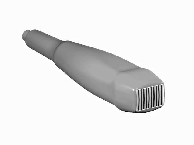
Figure 4: Image of an ultrasound probe for cardiology. The cable comes out from the top left and is connected to the ultrasound scanner. The front of the probe is about 20 x 15mm and only a few of the individual rod elements are shown here. In practice, there are between 48 and 128 such elements.
The ceramics has the property that it compresses and expands as the applied electrical voltage changes. This movement is transferred to the tissue. Conversely, mechanical pressure from the outside will generate an electrical voltage. The probe can thus be used both as transmitter and receiver by switching quickly between the transmitting and receiving modes. Imaging is based on the pulse-echo principle which is shared with radar and sonar. A short pulse is sent from the probe, propagates through the medium and returns a reflected pulse each time it meets a transition to a different tissue. The time it takes from sending the pulse until the echo is received by the probe gives a measure of the distance to the reflecting area.