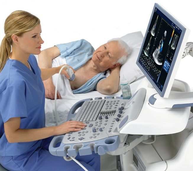
Figure 1: 3D ultrasound scanner for medicine, GE Vivid 7 Dimension (Used with permission from GE Healthcare).
In Norway, there is a large arena for technical and clinical development of medical ultrasound imaging. The company GE Vingmed Ultrasound in Horten develops, produces, and sells ultrasound instruments world-wide and is the "Center of Excellence" for cardiology ultrasound of GE Healthcare. There are about 200 employees in Norway and many outside of Norway. Figure 1 shows a typical ultrasound scanner. Vingmed was started by people from the technical-medial research environment at NTNU in Trondheim. It was the development of Doppler-based techniques in the 70s and 80s which provided the basis for Vingmed’s success. This was followed by the development of digital scanners with phased arrays in the 90s and new modes such as strain and real-time 3D in later years. Other companies include Medistim, which produces ultrasound equipment for quality control and documentation under cardiac surgery.
Figure 1: 3D ultrasound scanner for medicine, GE Vivid 7 Dimension (Used with permission from GE Healthcare).
The use of ultrasound, as well as research on clinical aspects of ultrasound, takes place at many hospitals in Norway. Research on the physical and technical aspects takes place at several universities. Furthermore, GE Healthcare (formerly Nycomed Amersham Imaging) had a group in Oslo which worked with the development of ultrasound contrast fluid, however this work ended in 2003.
There is also an active arena of ultrasound research in Denmark, especially at The Technical University of Denmark. They work closely with B-K Medical, which produces imaging systems specifically for urology and surgery, and which has almost 300 employees across the world.
A typical ultrasound heart image is shown in Figure 2. This is a 2-dimensional image which shows a slice through the heart. In this image, the brightness increases with the echo strength. The figure shows the heart as seen from the side (see Figure 3), and you can see the left ventricle to the left (depth 3.5-7.5 cm), the closed aortic valve and the aorta at the top right (depth 4-7 cm), the open mitral valve between the left atrium and ventricle in the middle of the image, and the left atrium at the bottom right (depth 8-11 cm). The walls of the heart are white in the image, as they give off a stronger echo than the blood in the chambers.
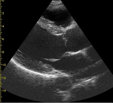
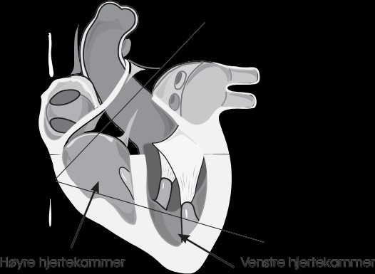
Figure 2 (left): 2-dimensional B-mode image from a person with good image quality, in the parasternal long axis view
Figure 3 (right): Anatomical image of the heart. The ultrasound image in Fig. 2 is taken from the cross-section which is marked.
These images are, as a rule, simpler to understand when they are shown as a time sequence rather than still images. One can immediately note that the walls of the heart and the blood in the chambers are not always distinctly separated, which makes e.g. automatic edge-detection difficult. This illustrates some of the challenges one faces with signal and image processing in ultrasound instruments.
The most important physical principles for ultrasound will be covered on this website. Furthermore, the most important ways in which these principles are utilized in scanners will be presented. The article is written for both medical students who wish to gain a better understanding of ultrasound imaging by learning background technology, and for physicists who wish to see how principles in acoustics are utilized in imaging. Formulas are shown in fact boxes so that those interested can study them as an extension of the main text.
The article starts with an overview of the most important physical principles which determine the resolution, and the depth of penetration. Then, the Doppler effect and effects of non-linear acoustics are discussed. The different methods of operation and types of images which are used for imaging of tissue and measurement of the velocity of blood flow is then covered. Next, we discuss the most important parts of an ultrasound scanner. Many types of probes and image formats have been developed. They are adaptated to limitations in size and accessibility presented by different parts of the body. Finally, we discuss safety with ultrasound imaging.
Professor Sverre Holm, Department of Informatics