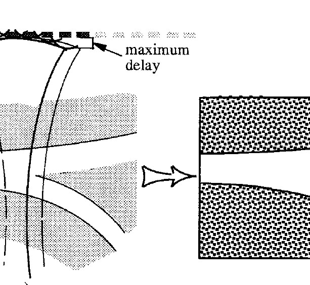
Figure 14: Linear scan. Left side shows the scanning situation where a probe is placed over the carotid artery, and the right side shows the resulting ultrasound image.
Figure 14: Linear scan. Left side shows the scanning situation where a probe is placed over the carotid artery, and the right side shows the resulting ultrasound image.
To get a good Doppler spectrum and good color Doppler, the beam must be angled relative to the blood flow. The beam must therefore be angled between 10-30° as shown in figure 15.
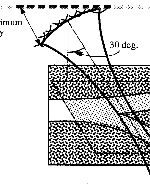
Figure 15: Linear scan with an angled beam for use in Doppler imaging.