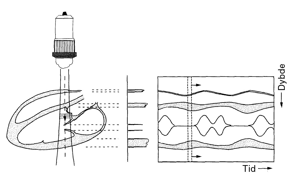
Figure 10: Illustration of M-mode. To the left, a heart with a beam that is fixed in space. To the right, the resulting image with time on the horizontal axis and distance from the probe on the vertical axis.
Figure 10: Illustration of M-mode. To the left, a heart with a beam that is fixed in space. To the right, the resulting image with time on the horizontal axis and distance from the probe on the vertical axis.
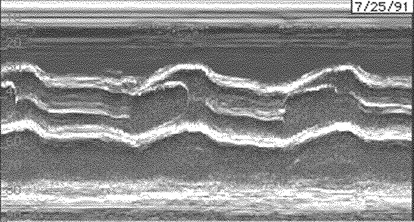
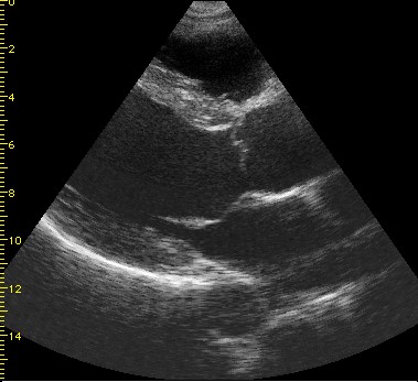
The images below are M-mode images (left) taken along a line in the B-mode image (right) through the aortic valve. The aorta and valve are visible. On the left, the valve is almost closed and the whole image depicts just over two heart beats.