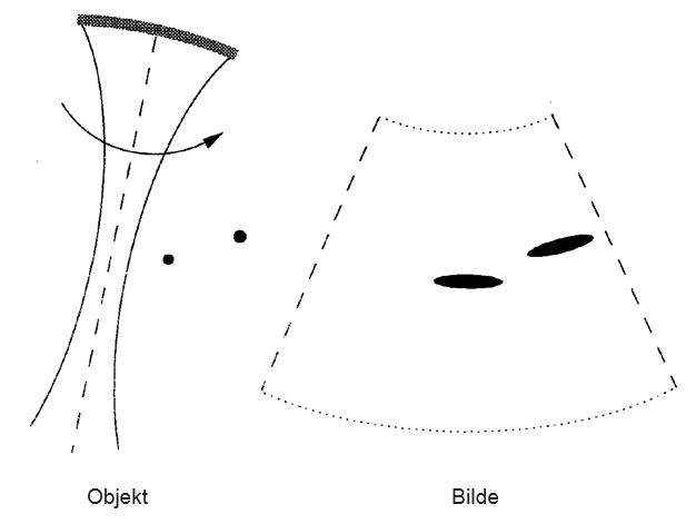
Figure 5: a) Scanning of two point-shaped objects, the probe with the sound wave to the left. b) The resulting ultrasound image. Notice that the lateral resolution is significantly worse than the radial resolution.
Figure 5: a) Scanning of two point-shaped objects, the probe with the sound wave to the left. b) The resulting ultrasound image. Notice that the lateral resolution is significantly worse than the radial resolution.
Since a short pulse has more frequency contents than a long one, it requires a larger bandwidth in the probe. In the 90s, new techniques and ceramic composite material made it possible to make probes with bandwidth 50-80% of the ultrasound frequency, and the trend is moving toward 100% relative bandwidth. Therefore, radial resolution is normally only a few wavelengths. Lateral resolution is dependent on the relationship between the wavelength and the size of the probe, it increases linearly with the distance from the probe. This is in contrast to radial resolution which is more or less independent of distance.
In contrast to, for example radar and most sonars, ultrasound displays objects in the nearfield of the probe. This is also a characteristic that is in common with optics, and therefore one needs to focus the beam. This is illustrated in figure 6. A probe with size D that is focused at a depth F is shown. The figure shows lateral beamwidth with depth towards the right. Focusing results in an optimal depth area, depth of field, as shown in the figure. In the ultrasound instrument, this focusing is done automatically upon signal reception. This is called dynamic focusing. Often, you can also adjust the transmitting focus manually.
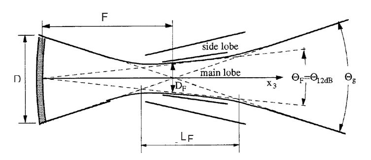
Figure 6: Beamwidth for a transducer with aperture D to the left in the picture. The focal point is at distance F and the depth of field is LF.
From what has been said about resolution, it should be an advantage to have the biggest possible probe in order to get the best possible lateral resolution. However, there are other limitations to take into account. In cardiology, the distance between the ribs restricts the size of the probe to less than 20 mm. Imaging of blood vessels in, for example, the throat and imaging of internal organs like the liver and kidneys do not have such restrictions. Other restrictions that can play a role are price of the equipment, as bigger probes generally require more electronical channels. For high quality equipment, physical limitations due to small aberrations in the tissue will also limit the benefit of bigger probes.