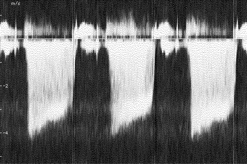
Figure 7: The Doppler spectrum recorded from the aortic valve through the heart apex. Positive speed is normal, negative represents a leakage.
The red blood cells are the most important spreaders of the Doppler shift. They give a quite weak signal compared to the echo of surrounding stationary tissue. This results in big requirements for dynamics in the design of the instruments. The Doppler frequency is normally chosen at the lower end of a probe's frequency bandwidth, for example, a 7.5 MHz probe uses 5-6 MHz as the Doppler frequency. With normal speed of blood flow, the difference between transmitted and received frequency appear in the audible range. It is therefore normal to play this on speakers while the velocity spectrum as a function of time is displayed on the screen of the ultrasound machine, see an example in figure 7.
Figure 7: The Doppler spectrum recorded from the aortic valve through the heart apex. Positive speed is normal, negative represents a leakage.