The probe both transmits and receives acoustic energy, and in continuous Doppler mode, it even does both simultaneously. It consists of many independent elemenets (up to ca 200) that can have a width down to 0.1 mm.
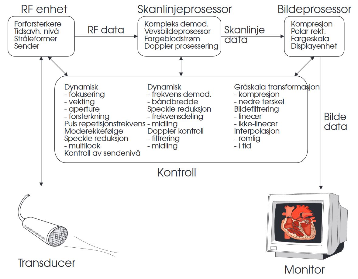
The RF (Radio Frequency) collection unit controls each element independently to achieve the desired deflection and focusing of the ultrasound beam. This unit consists of analog and digital electronics that combine the many signals from the probe to one output. This is done with the help of a beam former that dynamically delays certain channels with respect to others, and ensures that echo signals from each element in the probe are summed coherently. The beam former can be considered a signal processor that operates both in time and space domains. Today, it is made digitally, and the computational requirements are so heavy that it must be made from custom-made integrated circuits.
The scanline processor does signal processing in the time domain and extracts information from the RF-signal that has been collected. This processor is actually divided in three: one part to take out B- and M-mode data, one part for color Doppler and a spectral analyzer for spectral Doppler data. Implementation was previously done with the help of a signal processor, but today a standard PC can do this procedure.
The display consists of a software program that transforms scanline data to a format suitable for a screen. The most important operation is scan-conversion that is a transformation from a rectangular to a polar display format. In addition, it also compresses the amplitude and filters the image.
To control the whole scanner, there is also a control unit that is implemented as a software program. It ensures that all modules are controlled correctly and provide optimal extraction of information from the weak ultrasound signals.