In this format, 100-200 sound beams are transmitted at different angles as shown in the figure below and to the left. The advantage is a wide image through a small window. This format is used especially in cardiology (image to the right). Access is limited to the intercostal space. In the past one used mechanically tilted probes, but today electronically controlled probes (phased array) have taken over.
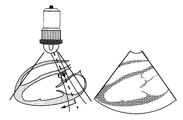
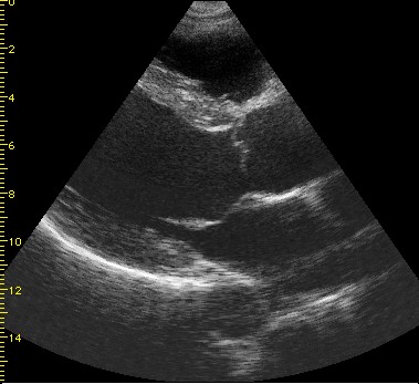
Figure 13: Sector scan format. Left side shows the scanning situation where a probe is held against the chest over the heart, and the right side shows the resulting ultrasound image.