The figure below shows a 2D image that is often called B-mode display, where B stands for Brightness. This is the standard image in ultrasound imaging. It shows the geometry of the organ and uses brightness to show the echo strength. It is based on transmitting and receiving on the same frequency, as in linear acoustics.

An image with more typical image quality is shown in figure 8. This image is taken from the apex of the heart, from the bottom up. This image is much more difficult to interpret as it has a much lower contrast.


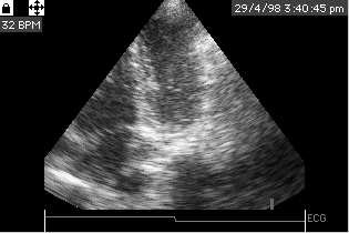
Figure 8: Image of a heart from the apex of the heart (apical) at 2.5 MHz based on linear acoustics.