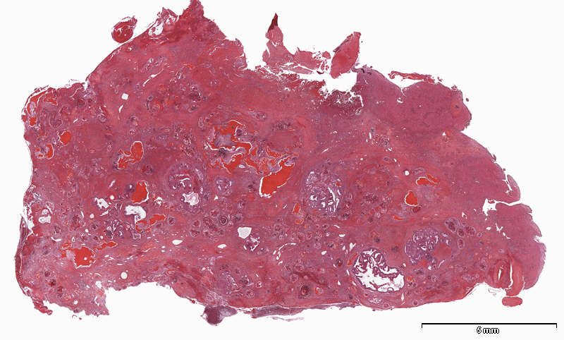
Patient history: 3 year-old girl with previously known enlarged liver and abdominal pain. Fine-needle aspiration showed hepatoblastoma. Having been treated with chemotherapy for tumor reduction, resection of the tumor and portions of the liver was undertaken.
This is a low power image of the resected specimen. You can see non-neoplastic liver tissue to the right side. The rest of the specimen shows a complex neoplastic tissue
Question: Indicate where in the this image you find epithelial and mesenchymal neoplastic tissue, respectively. |