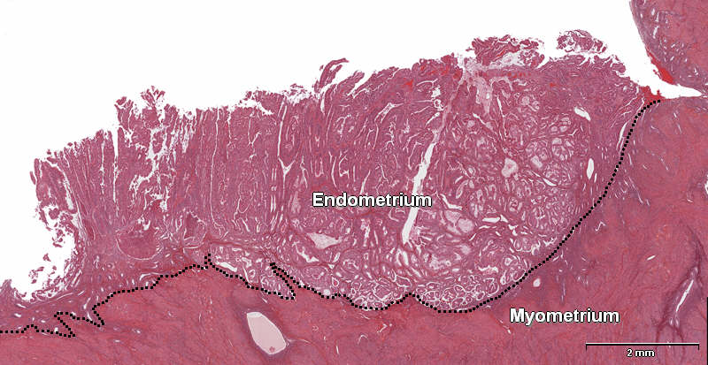
Patient history: 40 year-old female with irregular menstrual bleeding during the last 6 months. Cutterage showed adenocarsinoma upon histological examination. Admitted to hospital for removal of the uterus.
The image is a low power migrograph of endometrium and adjacent myometrium showing a well differentiated adenocarcinoma.
Question 1: Indicate the border between the two tissue components?
- see the line
Question 2: What kind of pathological change does the endometrium show?
- The endometrium is thicker than normal and displays atypical glands consistent with endometrial adenocarcinoma. Because you can see well developed glands, albeit atypical, the tumor is graded as a well differentiated adenocarcinoma. Because there is no infiltration into the myometrium, the tumor is classified as an adenocarcionoma in situ or intramucosal adenocarcinoma.
|