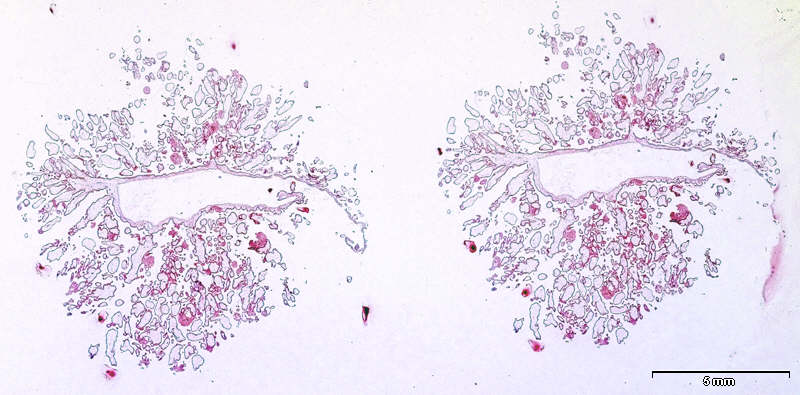
Two almost identical structures are seen. In the middle of each of them you have part of the chorionic cavity lined by the chorionic plate. The small spots of different sizes surrounding it are the chorionic villi. Note that the tissue sample has been folded during preparation. The chorionic plate is normally flat. |