 |
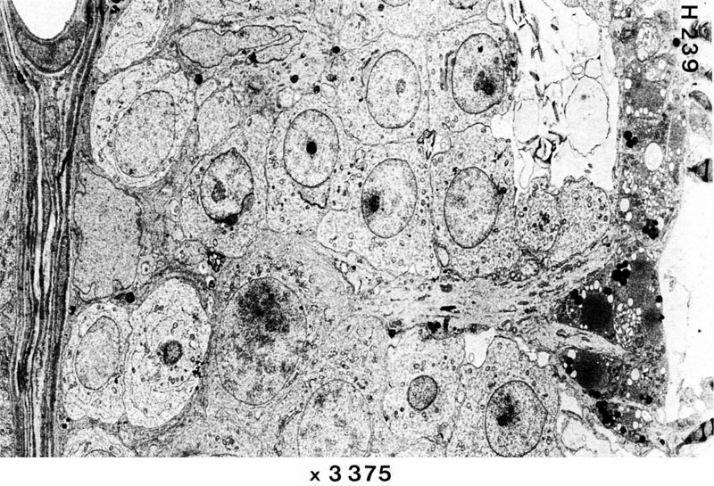
Low-power EM image through the wall of a seminiferous tubule. To the left you see layers of myopithelial cells and a small vessel (top). You also see cells in various stages of the spermatogenesis, from spermatogonia to spermatids. The further development of the spermatids to spermatozoa is not seen here. Note the irregular form of the Sertoli-cell nucleus. The cytoplasm of the Sertoli cells extend through the full thickness of the wall of the tubule. The dark material to the right (close to the lumen of the tubule) is cellular debris, which has been "thrown off" in the development of the spermatids to spermatozoa. |