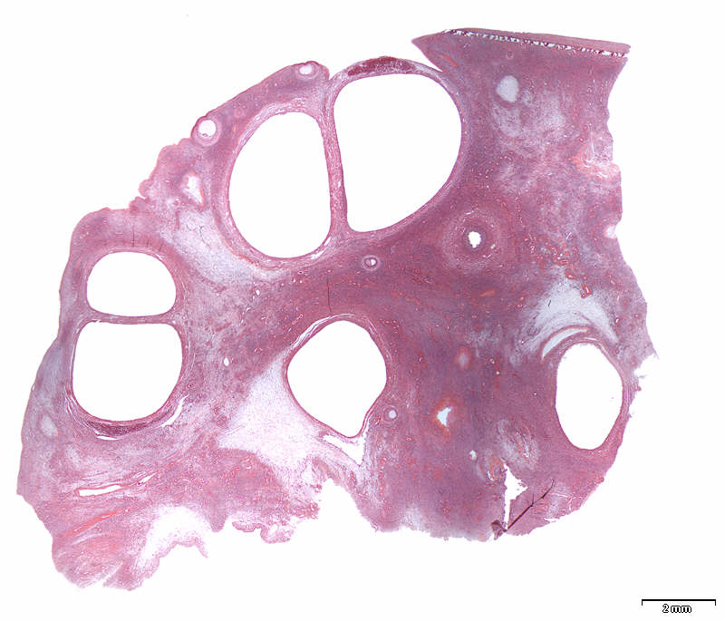
Patient history: 39 year-old female, undergoing in vitro fertilization (IVF) treatment for infertility. Ultrasonic examination showed bilateral polycystic ovaries. A wedge resection of both ovaries was performed and a section from the left ovary was taken for histological examination.
This image is a low power micrograph of the polycystic ovary. Find the cystically dialated follicles located mainly in the cortex, and the secondary follicles directly under the surface (see labelling by pressing ‘?’). |