Patient history: 63 year-old female with an abnormal tumor approx. 3 cm in diameter, severe ascites, discovered as a coincidental finding. Removal of the tumor and ovary was accomplished by a supravaginal uterus amputation. Parts of the omentum majus were resected.
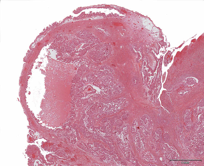
This low power micrograph of the ovary is showing the surface with its papillary tumor growth. The tumor tissue is seen infiltrating the ovarian stroma and cyst formation with partly papillary growth pattern.
Question 1: Why does this patient present with ascites? |