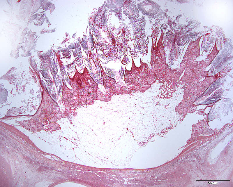
Patient history: 33 year-old female with a cyst in the right ovary, diagnosed upon gynecological examination. Right ovary and cyst were removed.
This is a low power micrograph showing parts of the inner cyst wall with verrucous epithelial lining with hyperkeratosis and fat tissue (see labeling by pressing '?').
Imagine the verrucous part seen in this image protruding into the cyst. The ovarian stroma is seen on the bottom of the image.
Question 1: Why is it then that we call this a dermoid cyst? |