Ubrukte filer
Hopp til navigering
Hopp til søk
Følgende filer eksisterer men er ikke innlagt på noen sider. Merk at andre sider kanskje lenker til en fil med en direkte lenke, så filen listes her selv om den faktisk er i bruk.
Nedenfor vises opptil 250 resultater fra og med nummer 21 til og med nummer 270.
Vis (forrige 250 | neste 250) (20 | 50 | 100 | 250 | 500)
 798px-Video konsept.gif 798 × 599; 51 KB
 100px-Tilt table videos.jpg 100 × 67; 4 KB
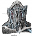 106px-Veins-of-the-neck-(Gray).jpg 106 × 120; 8 KB
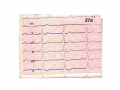 120px-Kids Ekg Karolinska.jpg 120 × 90; 5 KB
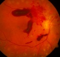 120px-Retinaleblødninger.jpg 120 × 114; 6 KB
 180px-Infusjonsoppheng.png 180 × 135; 44 KB
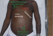 180px-Leverdempning-med-tekst.jpg 180 × 121; 10 KB
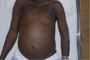 180px-Leverdempning.jpg 180 × 121; 9 KB
 180px-Presipitater.jpg 180 × 135; 10 KB
 180px-Schirmer.jpg 180 × 75; 3 KB
 180px-Trachea2.gif 180 × 150; 8 KB
 180px-Trommehinnen Gray.gif 180 × 131; 6 KB
 250px-Lungeundersøkelse.jpg 250 × 188; 17 KB
 250px-Standardjournal.png 250 × 188; 47 KB
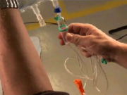
 250px-Venepunksjon.png 250 × 188; 82 KB
 300px-Logo UiO JournalWiki.png 300 × 56; 13 KB
 320px-T-celle-lymfom-ve-legg.jpg 320 × 240; 28 KB
 320px-Tavleogbrillekasse-screenshot.jpg 320 × 180; 31 KB
 400px-Forkammerreflekser.jpg 400 × 300; 43 KB
 450px-Eksoftalmometer.jpg 450 × 834; 78 KB
 500px-Bakteriellkeratitt.jpg 500 × 349; 61 KB
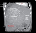
 514px-Levertumor1-m-tekst.jpg 514 × 480; 76 KB
 640px-Spaltelys.jpg 640 × 480; 90 KB
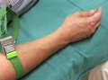
 640px-T-celle-lymfom-ve-arm.jpg 640 × 480; 69 KB
 640px-T-celle-lymfom-ve-legg.jpg 640 × 480; 97 KB
 70px-BHLR-voksne.gif 70 × 98; 3 KB
 70px-EKG AstraZeneca.jpg 70 × 70; 4 KB
 780px-Hypermetropi.jpg 780 × 599; 78 KB
 798px-Video konser.gif 798 × 599; 51 KB
 800px-Donders-screenshot.png 800 × 500; 523 KB
 800px-T-celle-lymfom-hø-fotsåle-nærbilde.jpg 800 × 600; 162 KB
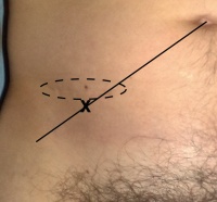
 800px-Ventralhernie laparoskopi.jpg 800 × 600; 124 KB
 800px-Ventralhernie stående.jpg 800 × 600; 118 KB
 800px-Øyestilling.jpg 800 × 588; 79 KB
 92px-Plantarrefleks.png 92 × 120; 20 KB
 94px-Pectus excavatum.jpg 94 × 120; 4 KB
 NormaloyeAndreas.jpg 2 360 × 1 472; 594 KB
 Oculocutaneous-albinism-LARGE.jpg 1 800 × 1 361; 396 KB
 Pas9hornhinnelinse2.jpg 3 062 × 2 269; 1 005 KB
 Pas 14 Protese3 nettside.jpg 3 110 × 2 238; 738 KB
 1024px-Fremre segment-screenshot.png 1 024 × 640; 784 KB
 1024px-Pupiller-screenshot.png 1 024 × 640; 791 KB
 120px-HLR BritishRedCross.png 120 × 145; 30 KB
 120px-Opening snap.PNG 120 × 68; 3 KB
 120px-Presipitater.jpg 120 × 90; 6 KB
 120px-Strabisme.jpg 120 × 77; 5 KB
 120px-Systolediastole.PNG 120 × 70; 3 KB
 120px-Tympanometri skjema.svg.png 120 × 80; 24 KB
 120px-Ventralhernie brokkport.jpg 120 × 90; 5 KB
 150px-Cardiology teaching package.jpg 150 × 113; 9 KB
 150px-ECG scribbles.jpg 150 × 113; 9 KB
 150px-Ecg wave maven.jpg 150 × 113; 9 KB
 161px-Aurikkel anatomi.JPG 161 × 239; 21 KB
 180px-Konjunktivitt.jpg 180 × 123; 13 KB
 180px-Kunstiglinse.jpg 180 × 127; 10 KB
 180px-Lungesinus.PNG 180 × 173; 27 KB
 180px-T-celle-lymfom-hø-ankel.jpg 180 × 135; 10 KB
 180px-T-celle-lymfom-ve-lår.jpg 180 × 135; 10 KB
 180px-Ventralhernie stående høyre.jpg 180 × 135; 9 KB
 180px-Øret anatomi oversikt Gray.jpg 180 × 159; 14 KB
 180px-Øyestilling.jpg 180 × 132; 8 KB
 202px-Venestuvning-eksempel1.jpg 202 × 174; 12 KB
 300px-Binasalhemianopsi.png 300 × 151; 55 KB
 300px-Papilleødem.jpg 300 × 225; 22 KB
 30px-Tips.svg.png 30 × 84; 4 KB
 320px-T-celle-lymfom-hø-arm-ulnart.jpg 320 × 240; 27 KB
 400px-Sagittal hode.gif 400 × 574; 60 KB
 400px-Trachea.gif 400 × 513; 23 KB
 500px-Myopi.jpg 500 × 372; 38 KB
 515px-McBurneyCloseup.jpg 515 × 480; 66 KB
 640px-Ventralhernie brokkport.jpg 640 × 480; 79 KB
 800px-Hirschbergstest.jpg 800 × 452; 91 KB
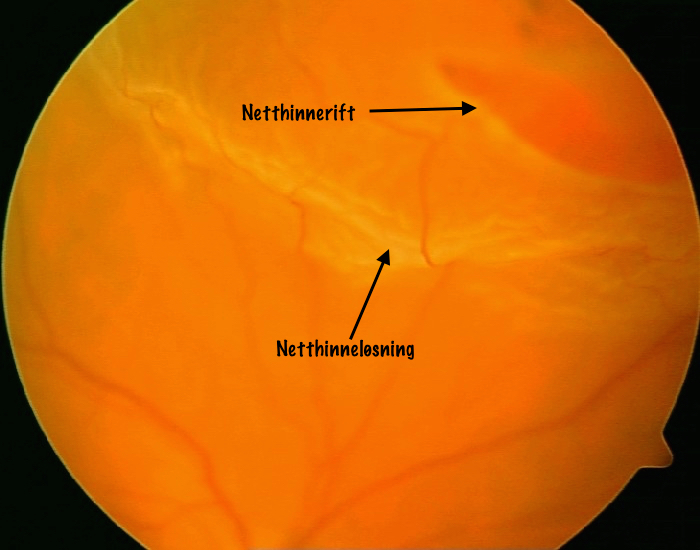 Netthinneløsning.jpg 700 × 550; 284 KB
 Normaloye1.jpg 1 024 × 768; 214 KB
 Schirmer.jpg 2 073 × 864; 151 KB
 0g4q732jcy8z113mak4j461dmc7vx2b.gif 600 × 422; 31 KB
 120px-Arcus costae.png 120 × 106; 20 KB
 120px-Bakteriellkeratitt.jpg 120 × 84; 7 KB
 120px-Deklive-ødemer1.jpg 120 × 81; 5 KB
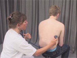
 120px-Littmann hjerte- og lungelyder.jpg 120 × 80; 5 KB
 120px-Lungevolumer.png 120 × 89; 8 KB
 120px-Lysvei.jpg 120 × 90; 4 KB
 120px-Ped-newborn.png 120 × 90; 22 KB
 120px-Venstrehomonymhemianopsi.png 120 × 60; 9 KB
 180px-Eksoftalmometer.jpg 180 × 334; 19 KB
 180px-Hypermetropi.jpg 180 × 138; 8 KB
 180px-Strabisme.jpg 180 × 115; 9 KB
 180px-T-celle-lymfom-hø-fotsåle.jpg 180 × 135; 11 KB
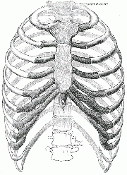 180px-Thorax bones.gif 180 × 248; 15 KB
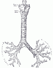 180px-Trachea.gif 180 × 231; 6 KB
 180px-Utspilt buk.jpg 180 × 268; 16 KB
 200px-McBurneyCloseup.jpg 200 × 186; 14 KB
 200px-Papilleødem.jpg 200 × 150; 11 KB
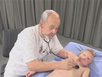 200px-Ped-cardiac-exam.png 200 × 150; 55 KB
 200px-Venestuvning-eksempel2.jpg 200 × 178; 12 KB
 250px-EKG University of Wisconsin Madison .jpg 250 × 188; 20 KB
 288px-Spaltelys.jpg 288 × 216; 24 KB
 300px-Bitemporalhemianopsi.png 300 × 151; 54 KB
 300px-Schirmer-test.jpg 300 × 180; 21 KB
 320px-T-celle-lymfom-ve-lår.jpg 320 × 240; 28 KB
 400px-Amotio.jpg 400 × 308; 24 KB
 400px-Lungelyder diagram.PNG 400 × 318; 69 KB
 450px-Linselux1.jpg 450 × 338; 48 KB
 50px-UniMarylandEKG.jpg 50 × 59; 3 KB
 600px-Papilleødem.jpg 600 × 450; 65 KB
 640px-Fremre segment-screenshot.png 640 × 400; 346 KB
 643px-Levertumor1-m-tekst.jpg 643 × 600; 108 KB
 643px-Levertumor1.jpg 643 × 600; 107 KB
 798px-UiO Med fak logo.png 798 × 114; 41 KB
 Ecgmadeeasy.jpg 312 × 475; 32 KB
 Pas 14 Protese nettside.jpg 2 470 × 993; 470 KB
 1078px-Covertest.jpg 1 078 × 1 024; 159 KB
 120px-1240sqyv2o94.25au6n.2.png 120 × 140; 28 KB
 120px-Auskultasjon screenshot.PNG 120 × 98; 18 KB
 120px-Exophthalmometer.jpg 120 × 41; 2 KB
 120px-Hodeomkrets.png 120 × 90; 22 KB
 120px-Ikterus farget2.jpg 120 × 90; 5 KB
 120px-Tilt table videos.jpg 120 × 80; 5 KB
 120px-Ventralhernie stående høyre.jpg 120 × 90; 5 KB
 150px-Sphygmomanometri.png 150 × 121; 12 KB
 180px-CSO video.jpg 180 × 135; 9 KB
 180px-Cor ausk.PNG 180 × 174; 37 KB
 180px-Inspeksjon av thorax.jpg 180 × 134; 11 KB
 180px-Lungegrenser ventralt.gif 180 × 171; 5 KB
 180px-Lysvei.jpg 180 × 135; 7 KB
 180px-Venestuvning-eksempel1.jpg 180 × 155; 10 KB
 180px-Øyetsomgivelser-screenshot.png 180 × 113; 31 KB
 200px-Blodtrykk.png 200 × 150; 56 KB
 200px-Ventralhernie fra innsiden.jpg 200 × 150; 15 KB
 278px-Spaltelys.jpg 278 × 209; 23 KB
 300px-Trachea2.png 300 × 251; 133 KB
 300px-Venstrehomonymhemianopsi.png 300 × 151; 53 KB
 320px-Bevegelighet-screenshot.png 320 × 200; 101 KB
 320px-Fremre segment-screenshot.png 320 × 200; 95 KB
 320px-Perkusjon screenshot.PNG 320 × 262; 90 KB
 402px-Utspilt buk.jpg 402 × 599; 58 KB
 42px-Tips.png 42 × 118; 4 KB
 57px-Tips.svg.png 57 × 160; 5 KB
 640px-Bitemporalhemianopsi.png 640 × 322; 240 KB
 640px-T-celle-lymfom-ve-hofte.jpg 640 × 480; 118 KB
 644px-McBurneyCloseup.jpg 644 × 600; 93 KB
 799px-Schirmer.jpg 799 × 333; 28 KB
 800px-Astigmatisme.jpg 800 × 547; 77 KB
 800px-T-celle-lymfom-hø-lår.jpg 800 × 600; 150 KB
 80px-404.png 80 × 80; 3 KB
 80px-BHLR-voksne.gif 80 × 112; 4 KB
 90px-Ventralhernie fra siden spontanreponert.jpg 90 × 120; 4 KB
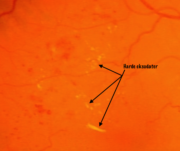 Harde eksudater.jpeg 683 × 573; 281 KB
 Logo UiO JournalWiki.png 2 786 × 520; 5,53 MB
 1024px-Hirschbergstest.jpg 1 024 × 579; 131 KB
 112px-Ventralhernie fra siden spontanreponert.jpg 112 × 149; 6 KB
 120px-11scocvu38w8.t1lqpt.2.png 120 × 17; 2 KB
 120px-404.png 120 × 120; 5 KB
 120px-Blodtrykk.png 120 × 90; 22 KB
 120px-Sphygmomanometri.png 120 × 96; 8 KB
 120px-Trachea2.png 120 × 100; 22 KB
 150px-AHLR AHA.png 150 × 97; 24 KB
 150px-HLR BritishRedCross.png 150 × 181; 46 KB
 160px-Perkusjon screenshot.PNG 160 × 131; 30 KB
 180px-Covertest.jpg 180 × 171; 11 KB
 180px-Hirschbergstest.jpg 180 × 102; 10 KB
 180px-Levertumor1-m-tekst.jpg 180 × 168; 13 KB
 180px-Seborrhoiskeksem.jpg 180 × 142; 13 KB
 180px-Synsfelt.png 180 × 136; 42 KB
 200px-Deklive-ødemer1.jpg 200 × 134; 11 KB
 200px-Indre øret Gray.gif 200 × 146; 9 KB
 250px-Abdomen.jpg 250 × 188; 16 KB
 300px-Auskultasjon screenshot.PNG 300 × 245; 80 KB
 300px-Splitting.PNG 300 × 170; 18 KB
 320px-Ventralhernie forfra spontanreponert.jpg 320 × 240; 22 KB
 500px-Astigmatisme.jpg 500 × 342; 37 KB
 500px-Spaltelys.jpg 500 × 375; 60 KB
 500px-Systolediastole.PNG 500 × 293; 37 KB
 506px-Covertest.jpg 506 × 480; 50 KB
 640px-Myopi.jpg 640 × 476; 55 KB
 640px-Ventralhernie forfra spontanreponert.jpg 640 × 480; 77 KB
 640px-Ventralhernie laparoskopi.jpg 640 × 480; 88 KB
 640px-Ventralhernie liggende under hoste.jpg 640 × 480; 65 KB
 75px-Synstavle.JPG 75 × 119; 5 KB
 800px-Forkammerreflekser.jpg 800 × 600; 129 KB
 800px-T-celle-lymfom-hø-arm-ulnart.jpg 800 × 600; 143 KB
 80px-Illustrasjon fra cardiovascular til PDA.jpg 80 × 34; 1 KB
 90px-Tilt table videos.jpg 90 × 60; 4 KB
 947px-Pupillarplanet.jpg 947 × 1 024; 172 KB
 Pas10neglelimfluorescein.jpg 1 442 × 936; 586 KB
 Utenfor Pensum.jpg 23 × 21; 4 KB
 100px-Schirmer-test.jpg 100 × 60; 4 KB
 119px-Subkonjunktival blødning.jpg 119 × 120; 7 KB
 120px-11scobo0u3zg.9i5ft6.2.png 120 × 17; 2 KB
 120px-Illustrasjon clinical exam.jpg 120 × 110; 8 KB
 120px-Perkusjon screenshot.PNG 120 × 98; 19 KB
 120px-Spaltelys.jpg 120 × 90; 6 KB
 120px-Subkonjunktival blødning.jpg 120 × 83; 5 KB
 120px-Venepunksjon.png 120 × 90; 22 KB
 120px-ؘyestilling.jpg 120 × 88; 4 KB
 180px-Donders-screenshot.png 180 × 113; 34 KB
 180px-Epiglottis-(Gray).png 180 × 175; 49 KB
 180px-Indre øret Gray.gif 180 × 131; 8 KB
 180px-Linselux1.jpg 180 × 135; 9 KB
 180px-T-celle-lymfom-hø-legg.jpg 180 × 135; 11 KB
 200px-Tavleogbrillekasse-screenshot.jpg 200 × 113; 14 KB
 200px-Thorax muscles.png 200 × 194; 68 KB
 200px-Uncovertest.jpg 200 × 219; 12 KB
 200px-Ventralhernie forfra spontanreponert.jpg 200 × 150; 10 KB
 200px-Øret anatomi oversikt Gray.jpg 200 × 177; 17 KB
 250px-Auskultasjon screenshot.PNG 250 × 205; 61 KB
 250px-Papilleødem.jpg 250 × 188; 16 KB
 250px-Ped-one-year.png 250 × 187; 85 KB
 300px-Aortastenose.PNG 300 × 170; 20 KB
 320px-T-celle-lymfom-hø-arm-radialt.jpg 320 × 240; 27 KB
 320px-UiO Med fak logo.png 320 × 46; 10 KB
 350px-Exophthalmometer.jpg 350 × 121; 10 KB
 400px-Thorax bones.gif 400 × 550; 67 KB
 500px-Krysningsfenomener.jpg 500 × 455; 50 KB
 631px-Covertest.jpg 631 × 599; 71 KB
 639px-Video konsept.gif 639 × 480; 35 KB
 640px-T-celle-lymfom-hø-legg.jpg 640 × 480; 109 KB
 79px-Ecgmadeeasy.jpg 79 × 120; 7 KB
 800px-Evertering-screenshot.png 800 × 500; 542 KB
 800px-Tympanometri skjema.svg.png 800 × 533; 289 KB
 Colobom2.jpg 415 × 358; 88 KB
 Pas 12 Ektropion enkelt forfra.jpg 1 065 × 750; 199 KB
 100px-404.png 100 × 100; 3 KB
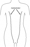 100px-Melkelister.png 100 × 154; 10 KB
 1024px-Donders-screenshot.png 1 024 × 640; 792 KB
 1024px-Myopi.jpg 1 024 × 761; 115 KB
 113px-Ventralhernie fra siden spontanreponert.jpg 113 × 151; 6 KB
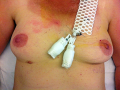 120px-Aksessorisk-brystvorte1.png 120 × 90; 23 KB
 120px-Levertumor1-m-tekst.jpg 120 × 112; 7 KB
 120px-Lungeundersøkelse.jpg 120 × 90; 6 KB
 120px-PVK-stase.jpg 120 × 89; 6 KB
 120px-Video konser.gif 120 × 90; 2 KB
 150px-Littmann hjerte- og lungelyder.jpg 150 × 100; 7 KB
 150px-Sigve holmen EKG.jpg 150 × 88; 5 KB
 150px-The auscultation assistant.jpg 150 × 75; 7 KB
 180px-Ejeksjonsklikk.png 180 × 102; 7 KB
 180px-HLR BritishRedCross.png 180 × 217; 64 KB
 180px-Keratoconus.jpg 180 × 273; 18 KB
 180px-Lungelyder diagram.PNG 180 × 143; 17 KB
 180px-Schirmer-test.jpg 180 × 108; 9 KB
 180px-Systolediastole.PNG 180 × 105; 6 KB
 180px-Thorax muscles.gif 180 × 174; 9 KB
 200px-Inspeksjon av thorax.jpg 200 × 149; 13 KB
 250px-McBurneyCloseup.jpg 250 × 233; 20 KB
 300px-Bakterieprøve-screenshot.png 300 × 188; 88 KB
 300px-Hirschbergstest.jpg 300 × 170; 21 KB
 312px-Hypermetropi.jpg 312 × 240; 19 KB
 320px-Bitemporalhemianopsi.png 320 × 161; 61 KB
 320px-Lungevolumer.jpg 320 × 200; 15 KB
 320px-Video konsept.gif 320 × 240; 11 KB


.jpg)


.png)


